When treating sciatica, it’s very important to understand the anatomy in the spine and pelvis because knowing the source of the problem will help you to follow the correct treatment plan for best results.
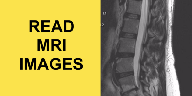
In this video I show you the anatomy of the lumbar spine as seen on an MRI image and I go over what your doctor looks for when making a diagnosis in the low back.
An MRI shows the integrity of your discs and shows soft tissue injuries that can’t be seen on an x-ray study.
For this reasons MRI’s are best when looking for MRI pathologies such as disc herniations that may be the cause of sciatica or simply low back pain that is sharp in nature.
WHAT TO LOOK FOR WHEN YOU READ AN MRI
1. Color of the disc
In an MRI image, the intervertebral disc is shown as white due to the fact that it contains water. A normal disc will show as white, whereas a degenerated disc or herniated disc will show up as black.
2. Spacing in between vertebrae
The discs act as shock absorbers and help to separate vertebrae from each other. This separation is what creates the opening where nerves come out of. If the disc space is narrowed, it indicates that the disc is either degenerating or herniated and can cause pinched nerves.
3. Posterior border of a disc
Because a disc is weaker at its posterior border, the majority of herniations are posterior. Since the spinal cord and nerve roots lie posterior to the disc, these can become compressed when a disc herniates. On an MRI image we look for a rounded posterior border to indicate a disc herniation.
Those are 3 things your doctor looks for when reading an MRI of the spine and when looking for disc herniations.


